HUMAN VERTEBRAL COLUMN
The human vertebral column is commonly referred to as the backbone or spine.
For the purpose of understanding proper anatomical alignment in relation to proper physical structure required for efficient execution of movement, the human vertebral column is the single most important anatomical structure. If we wish to gain any genuine comprehension of proper skeletal alignment, an adequate knowledge of the human vertebral column, it's anatomical structure, and how it functions is imperative.
STRUCTURE OF THE HUMAN VERTEBRAL COLUMN
The human vertebral column consists of approximately 33 vertebrae. These are divided into different regions, which correspond to the curves of the spinal column. These regions are called:
- Cervical,
- Thoracic,
- Lumbar,
- Sacrum,
- Coccyx.
The articulating vertebrae are named according to their region of the spine. There are seven cervical vertebrae, twelve thoracic vertebrae and five lumbar vertebrae. The number of vertebrae in a region can vary but overall the number remains the same. The number of those in the cervical region rarely changes.
INDIVIDUAL VERTEBRA
The vertebrae of the cervical, thoracic, and lumbar sections are independent articulating bones. The vertebrae of the sacrum and coccyx are usually fused and unable to move independently. Two special vertebrae are the atlas and axis, on which the cranium rests.
A typical vertebra consists of two parts: the vertebral body (anterior) and the vertebral arch (posterior). Together, these enclose the vertebral foramen, which contains the spinal cord. Because the spinal cord ends in the lumbar spine, and the sacrum and coccyx are fused, they do not contain a central foramen.
Above and below each vertebra are Zygapophyseal joints. These joints restrict the range of possible movement. In between each pair of vertebrae are two small holes called intervertebral foramina. Spinal nerves branch from the spinal cord and extend into the body through these holes.
Individual vertebra are named according to their region and position. From top to bottom, the vertebrae are:
- Cervical: 7 vertebrae (C1–C7)
- Thoracic: 12 vertebrae (T1–T12)
- Lumbar: 5 vertebrae (L1–L5)
- Sacrum: 5 (fused) vertebrae (S1–S5)
- Coccyx: 4 (3–5) (fused) vertebrae (tail bone)
INTERVERTEBRAL DISCS
Intervertebral discs are located between adjacent vertebrae in the vertebral column. Each disc forms a fibrocartilaginous joint. These joints allow for slight movement of individual vertebra and act as a ligament binding vertebrae together. Their function as shock absorbers in the human vertebral column is crucial.
STRUCTURE OF INTERVERTEBRAL DISCS
There is one disc between each pair of vertebrae, except for the first cervical segment, the atlas. The atlas is a ring around the roughly cone-shaped extension of the axis (second cervical segment). The axis acts as a post around which the atlas can rotate, allowing the neck to swivel.
There are 23 discs in the human vertebral column:
- Cervical: 6 in the neck,
- Thoracic:12 in the middle back,
- Lumbar: 5 in the lower back.
FUNCTION OF INTERVERTEBRAL DISCS
The intervertebral discs function to separate individual vertebra from each other. Intervertebral discs serve as a shock-absorbing surface. Each disc functions to distribute hydraulic pressure in all directions within each intervertebral disc under compressive loads. The segmented structure of the human vertebral column causes intervertebral discs to function as a hydraulic chain. Intervertebral discs degenerate with age.
FOUR CURVATURES OF THE HUMAN VERTEBRAL COLUMN
The four curvatures of the human vertebral column consist of the:
- convex cervical curvature
- concave thoracic curvature
- convex lumbar curvature
- concave sacral curvature
CONVEX CERVICAL CURVATURE
Generally, the cervical vertebrae exhibit a convex curvature to the anterior. This curvature begins at the second cervical vertebra and ends at the middle of the second thoracic vertebra. This is the least marked of the four curves.
CONCAVE THORACIC CURVATURE
Generally, the thoracic vertebrae exhibit a concave curvature to the anterior. This curvature begins at the middle of the second thoracic vertebra and ends at the middle of the twelfth thoracic vertebra. This curve is known as a kyphotic curve.
CONVEX LUMBAR CURVATURE
Generally, the lumbar vertebrae exhibit a convex curvature to the anterior. This curvature begins at the middle of the last thoracic vertebra and ends at the sacrovertebral angle. The convexity of the lower three vertebrae are much greater than that of the upper two. This is described as a lordotic curve. Typically, the lumbar curve is more marked in females than in males.
CONCAVE SACRAL CURVATURE
Generally, the sacral vertebrae exhibit a concave curvature to the anterior. This curvature begins at the sacrovertebral articulation and ends at the termination point of the coccyx. Its concavity is directed downward and forward.
SIGNIFICANCE OF THESE FOUR CURVATURES OF THE HUMAN VERTEBRAL COLUMN
It's absolutely imperative to note the thoracic and sacral curves are primary curves! They are present in fetal development.
The cervical and lumbar curves are compensatory and develop after birth! The cervical curve forms when the infant is able to hold up its head, at approximately three or four months of age, and to sit upright, at approximately nine months of age. The lumbar curve forms when the child begins to walk, at approximately twelve to eighteen months of age.
SINGLE GREATEST KEY TO PROPER PHYSICAL STRUCTURE
This means that concave curvature of the human vertebral column to the anterior is the result of natural anatomically correct structural design! Conversely, convex curvature of the human vertebral column to the anterior is the result of developmental pathology! This is the single greatest key to unlocking the mystery of proper structure! Understanding this provides a scientific answer to the question of how to properly achieve anatomically correct alignment of our physical structure as a means to facilitate effective, efficient, and nearly effortless movement!
Anatomically correct alignment of the human vertebral column is the primary key to proper physical structure. It's the primary key to nearly effortless movement, balancing pressure, generating force, increased acceleration, improved stability, greater flexibility, gaining leverage, and authentic mastery of technique. It's the primary key to dominating a physically larger and stronger adversary.
Genuine understanding of this key is the first step toward reliance on superior technique rather than athleticism and brute force. It's the first step to refrain from struggling with or "muscling" an opponent. It's the first step toward flowing with the winds of his hatred and leading him to the very destruction he seeks. It's the first step toward graceful and elegant movement in the midst of violent conflict. It's the first step toward truly dancing harmoniously with an assailant.
ANATOMICALLY CORRECT ALIGNMENT OF THE HUMAN VERTEBRAL COLUMN
The most common error among practitioners of various martial arts is an assumption that to achieve anatomically correct alignment of the human vertebral column results in flattening out, both convex and concave, curvatures into a straight vertical line. While this certainly yields significant improvement of structural integrity by eliminating the pathological convex curvature of the human vertebral column to the anterior, it is not anatomically correct. Straightening the spine may be considered correct posture socially or culturally, but it is incorrect anatomically.
Anatomically correct alignment of the human vertebral column creates a general concave curvature to the anterior!
 |
Notice that concave curvature of Lee's vertebral column to the anterior and the pelvic tilt.
|
SUPERIORITY OF CONCAVE VERTEBRAL CURVATURE
By eliminating only the two developmentally pathological convex curvatures of the cervical and lumber vertebrae, the human vertebral column creates a general concave arc from cranium to coccyx. A cursory study of architectural engineering will confirm that, all else being equal, a geometric arc or arch possesses greater structural integrity than a straight column or beam. A brief study of physics will also confirm that, all else being equal, a sphere possesses greater structural integrity than a cube. Even simple logic suggests that, when directly facing an adversary or an incoming force, a concave curvature of the human vertebral column is preferable. This theory is easily tested and confirmed by experience.
 |
| Anatomically correct alignment of the human vertebral column. |
ADVANTAGES OF CONCAVE VERTEBRAL CURVATURE
The immediate and direct advantages of concave vertebral curvature are:
- increased structural integrity,
- elimination of unnecessary muscle tension,
- elimination of unnecessary pressure on vertebrae,
- elimination of unnecessary pressure on intervertebral discs,
- proper protective positioning of cranium, eyes, and nose,
- proper protective positioning of cervical-cranial joint, mandible, and esophagus,
- proper protective positioning of shoulders,
- proper protective positioning of thorax,
- proper protective positioning of elbows,
- proper protective positioning of abdomen,
- proper protective positioning and support of internal organs,
- proper protective positioning of pelvis,
- proper protective positioning of knee joints,
- prevents knee joints from improper locking,
- allows for proper balance,
- allows for rapid and nearly effortless transfer of weight,
- allows for rapid and nearly effortless movement of limbs.
Implementation of concave vertebral curvature results in a multitude of indirect or secondary advantages as a consequence of the direct and primary advantages listed above. Generally, adopting such a posture will improve mobility, flexibility, ability to manage external physical pressure, ability to generate pressure, ability to reduce injury, and increase speed of movement. Additionally, implementation of concave vertebral curvature can, over time, result in numerous long term health benefits.
UNIQUE STRUCTURAL QUALITIES AND FUNCTION OF THE HUMAN VERTEBRAL BRIDGE
The human vertebral column possesses rather unique structural qualities. Unlike any other bones of the human skeletal system, the human vertebral column is not a single unit. The physical structure of the human vertebral column is segmented. The physical structure of the human vertebral column is not a straight solitary unit like the major bones of our limbs, such as the tibia, fibula, femur, radius, ulna, and humerus.
The human vertebral column features unique structural qualities, because it is anatomically designed for a unique and specific purpose. The human vertebral column functions differently from other bones of the human skeletal system. Furthermore, we must not be deceived by its nomenclature.
The vertebral column is not a column! Nor is it designed to function as such! In fact, it is a vertebral bridge and is designed to function as such.
Like it or not, serious study of human anatomy makes it painfully obvious that we are not physically designed to walk as completely vertical and upright as we presently do. Based on the anatomical structure of the human vertebral column, the anatomical structure of our musculature, the manner of physiology in which our organs are attached to and hang from our vertebral column, it is undeniable that we are anatomically designed to be knuckle draggers.
Our spines are a heritage from distant ancestors, men who carried themselves at a significantly greater horizontal angle.
Our spine is structurally designed to function like a flexible suspension bridge in support and protection of internal organs, joints, musculature, nerves, and other tissue. Over time, human behavior has forced our spine to function as a weight bearing column, placing it under great stress that's likely to cause eventual back injury and pain.
STRUCTURE AND FUNCTION OF AN ARCHED VERTEBRAL BRIDGE
The structural arch features prominently in architectural bridge designs and with good reason. Its semicircular structure elegantly distributes (redirects) pressure (force) through its entire structural form and diverts that pressure (force) onto its abutments (legs), which are the components (columns) of the bridge that directly transfer pressure (force) to the foundation (earth). Pressure (force) on the structure of an arch bridge itself is virtually negligible. The natural geometric curve of the arch itself causes pressure (force) to dissipate (redirect). This greatly reduces the effects of pressure (force) on both exterior (posterior) and interior (anterior) surfaces of the structure (arch bridge) itself.
In a naturally relaxed state, our spine forms the anatomically correct form and function of an arched vertebral bridge.
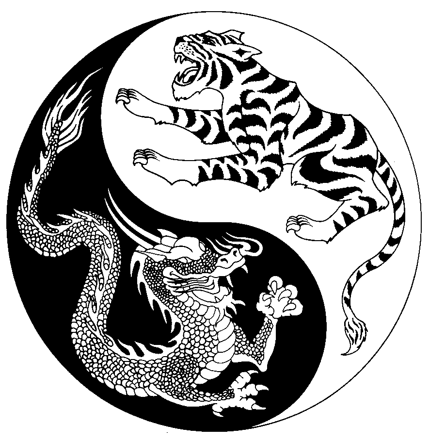
DIAGRAM OF SUPREME ULTIMATE
The diagram of supreme ultimate (太極 圖 - taiji tu) is a symbolic representation for the principle of
seemingly opposing forces acting harmoniously in relation to the function of any unified system. Additionally, it is intended to illustrate the illusion of duality. The object being to understand that all things possess a dual nature. This dual nature manifests itself as polar extremes. These polar extremes are represented by yin and yang.
DUAL NATURE OF THE STRUCTURAL ARCH
The concave interior (anterior) structure of the arch is yin in nature and designed to
receive pressure (force). The convex exterior (posterior) structure of the arch is yang in nature and designed to
repel pressure (force).
A profound understanding of these principles clearly confirms the correct logic of implementing an anatomically correct alignment of an anterior concave vertebral bridge as the proper physical structure in order to manage incoming pressure from any external source and in order to generate internal pressure intended to be applied to any external structure.
STRUCTURE AND FUNCTION OF THE FETAL POSITION
To further illustrate the structure and function of an arched vertebral bridge, study the structure of the fetal position.
It is clearly designed as a natural and anatomically correct structure for protective defense. The anatomically correct convex posterior surface of the vertebral bridge is positioned toward the external environment to repel incoming pressure. Simultaneously, the anatomically correct concave anterior surface of the vertebral bridge is positioned toward the internal environment to receive incoming pressure. Notice the protective positioning of cranium, eyes, nose, mandible, esophagus, shoulders, thorax, elbows, abdomen, internal organs, and pelvis.
Regardless of positioning of the legs and feet, regardless of orientation, standing upright, inverted upside down, or laying down, the principles hold true. That is precisely what determines the validity of a principle. It is universally true regardless of circumstance.
CONCLUSION
With a profound understanding concerning the fundamental structure and function of the anatomically correct alignment of our spine, we can proceed toward understanding the arched vertebral bridge principle and learning how to correctly implement this principle in the execution of establishing proper physical structure.


